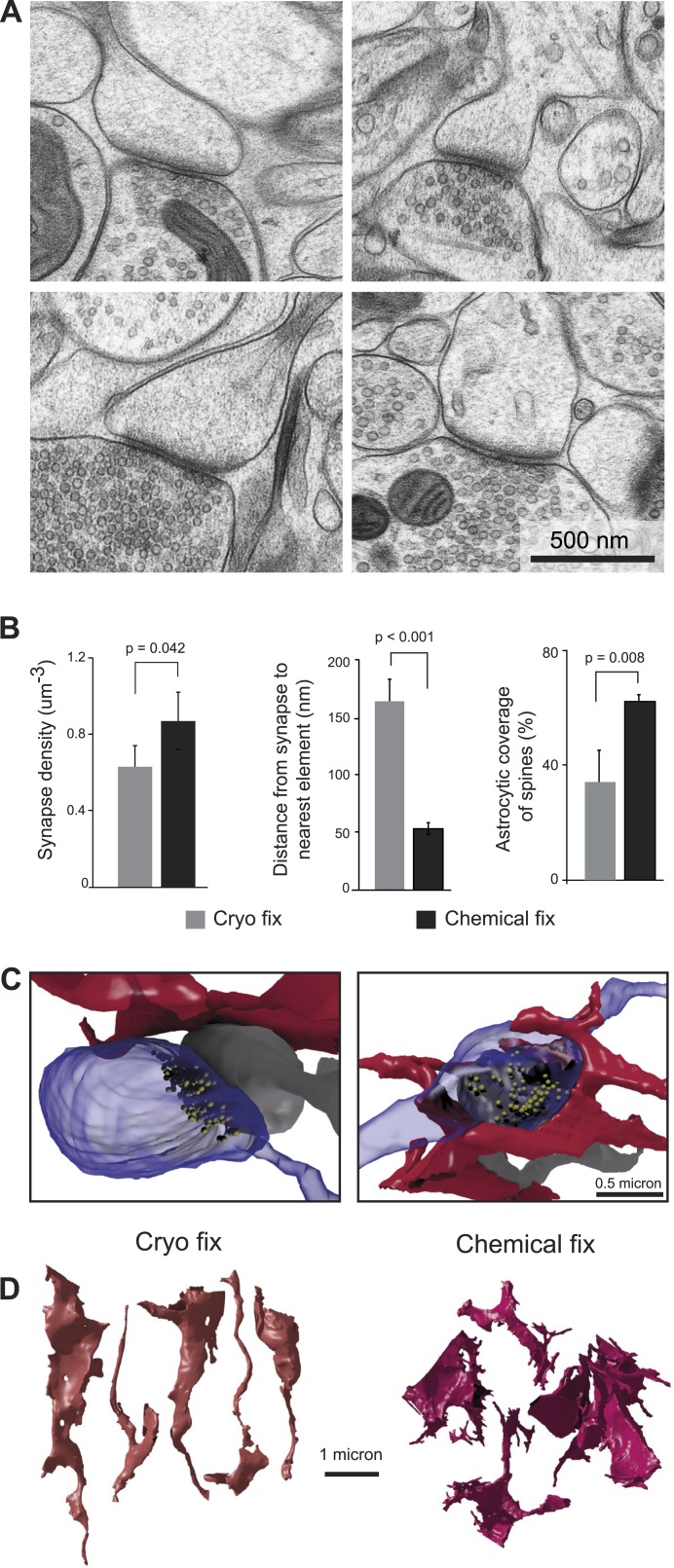
Figure 2. Cryo fixation reveals a larger peri-synaptic space and reduced astrocytic coverage.
(A) Cryo-fixed neuropil shows synaptic contacts with large amounts of surrounding extracellular space. (B) Synaptic density measurements show the chemically fixed neuropil to have 38% more synapses (left graph, p < 0.05, one way ANOVA). Dendritic spine synapses (presumed glutamatergic) show greater distances between the edge of the contact zone and the nearest membrane compared with chemical fixation (middle graph, p < 0.001, unpaired Student's t-test). Cryo-fixed synapses show less astrocytic coverage (right graph, p < 0.01; one way ANOVA). (C) Reconstructions from serial electron microscope images, of axonal boutons (blue) synapsing with dendritic spines (grey), show the astrocytic processes in the near vicinity (red). In the cryo-fixed synapse (left), the astrocytic process is not squeezed close to the synaptic contact (indicated with vesicles in yellow). In the chemically fixed example (right), the astrocyte tightly surrounds the synapse, where the vesicle-filled axonal bouton contacts the spine behind it. (D) Astrocytic processes reconstructed from serial FIBSEM images using the ilastik software (www.ilastik.org) show that chemically fixed astrocytic processes (right) have a more elaborate morphology with small processes extending from the flattened lamellae compared with the less complex structure of cryo-fixed astrocytes (left).
DOI: http://dx.doi.org/10.7554/eLife.05793.007

