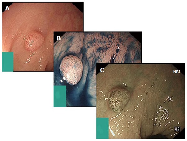
Figure 3.

Digital chromoendoscopy. A: Represents sessile adenoma seen in standard white light; B: Shows the same adenoma after the use of indigo carmine applied for chromoendoscopy; C: Shows further assessment of the adenoma using narrow band imaging (NBI).
