
Figure 4.
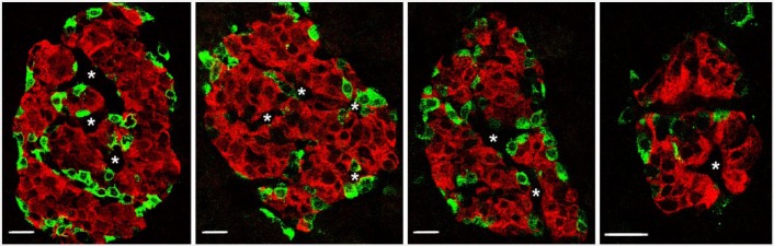
Islets in pancreas from donor H0021 immunofluorescently stained for insulin (red) and non-β-cells (glucagon and somatostatin; green) show clear clustering of β-cells with surrounding non-β-cells. Additionally, apparent vascular channels (white asterisks) are seen penetrating in the β-cell clusters. Scale, 20 µm.