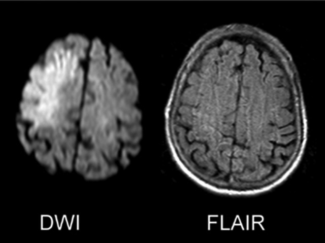
Figure 2.

The DWI/FLAIR mismatch. These 2 axial images of the brain at a level just above the lateral ventricles represent the so-called DWI/FLAIR mismatch that can be seen in the early hours after symptom onset when DWI (left) hyperintensity—which can arise in minutes from symptom onset—occurs in the absence of T2-based FLAIR (right) hyperintensity, which takes 3 to 6 hours to develop. DWI indicates diffusion-weighted imaging; FLAIR, fluid attenuated inversion recovery.
