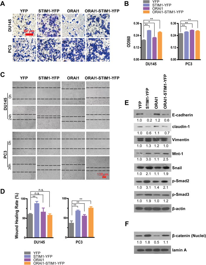
Figure 5. Overexpression of STIM1 and/or ORAI1 promotes the migration of DU145 and PC3 cells.
A. Representative crystal violet staining for DU145 and PC3 cells that migrated and attached to the bottom of transwell filters after 24 h of treatment. B. Statistical analysis of crystal violet staining intensity measured as the absorbance at 560 nm, (n = 3 for DU145 cells, n = 5 for PC3 cells). C. Wound-healing assays of cell motility in DU145 and PC3 cells, and representative images obtained at 0 and 24 or 36 h after scratching. The dashed line indicated the edge of the wound. D. Statistical analysis of the wound-healing rates of DU145 and PC3 cells, (n = 5 for DU145 cells, n = 3 for PC3 cells). E. Western blotting analysis of E-cadherin, claudin-1, Vimentin, Wnt-1, Snail, p-Smad2 and p-Smad3 in DU145 cells. F. Western blotting analysis of nuclear β-catenin in DU145 cells.

