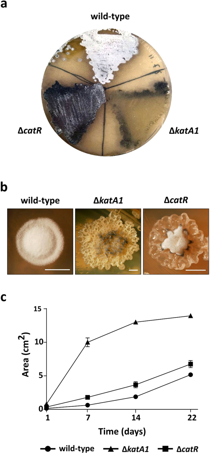
Figure 1. Morphological phenotypes and mycelium proliferation assay of S. natalensis wild-type, ΔkatA1 and ΔcatR strains grown in R5 solid medium.

(a) Photographs of mycelium lawns at 72 h. (b) Photographs of isolated colonies at 72 h. Scale bar: 2,50 mm. Photographs by Tiago Beites. (c) Mycelium proliferation rate in R5 solid medium. 10 μl drops of liquid cultures grown to an OD600nm of 4–5 were placed in the middle of R5 plates and mycelium proliferation was measured for 22 days. R5 plates were photographed and the mycelium area was determined using the measure function of the ImageJ software. Results are representative of three independent experiments.
