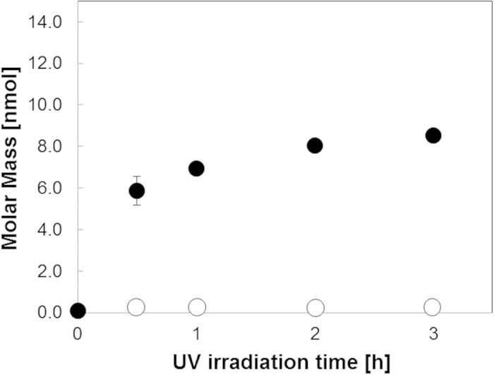
Figure 2. Amount of peptide cleaved versus UV (365 nm) irradiation time.

Peptides on the membrane spots (n = 3) were cleaved by UV irradiation (365 nm). Black circles denote Fluorescein-GABA-RRRRRRRR-Photo-cleavable linker, which can be cleaved by UV. White circles denote Fluorescein-GABA-RRRRRRRR-11-aminoundecanoic acid, which cannot be cleaved by UV (negative control). Peptide concentration was determined by measuring the amount of fluorescence. The standard curve of the purified Fluorescein-GABA-RRRRRRRR was used to determine the concentration of the cleaved peptide.
