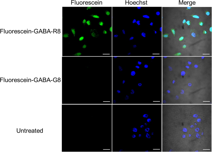
Figure 3. Internalization of the detached peptide by HeLa cells.
Confocal microscopy analysis of HeLa cells was conducted after incubating the cells at 37 °C for 3 h with Fluorescein-GABA-R8, Fluorescein-GABA-G8 as a negative control, and untreated control. About 1.5 × 10⁵ HeLa cells were seeded in 35-mm glass-bottom dish 24 h before the experiment. Scale bar = 30 μm.

