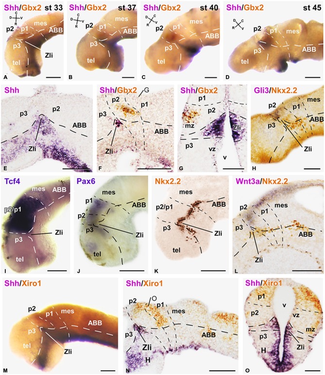
Figure 1.
Expression of early markers of the diencephalic prepatterning and formation of the Zli. Microphotographs of lateral views of the forebrain in whole mounts labeled for double in situ hybridization (ISH) to reveal Shh (purple) and Gbx2 (orange) at the embryonic stages indicated (A–D); the orientation is indicated in each panel to highlight the dorsoventral and rostrocaudal change of the axis through the thalamus at each stage. (E–O): Microphotographs of whole mounts (I,J,M) and sagittal (E,F,H,K,L,N) or transverse (G,O) sections of embryos at stages 33/34. The photographs correspond to single ISH (E,I,J), single immunohistochemistry (IHC; K), and combinations of double ISH (F,G,M–O) or combined ISH and IHC (H,L). The markers labeled are indicated in the upper left of each photograph. All images are oriented following the same standard: dorsal is upwards in transverse and sagittal sections, and rostral is to the left in sagittal sections. The neuromeric boundaries and main brain subdivisions are indicated to assist in the precise localization of the labeling. The levels of the transverse sections (G,O) are indicated in photographs (F,N), respectively. Scale bars = 100 μm (E,F,L–N), 50 μm (A–D,G–K,O). See list for abbreviations.

