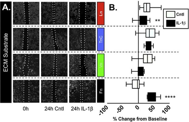
Fig. 1. Astrocytic response to scratch response and IL-1β is determined by ECM substrate.
(A) Mixed glial cultures grown on cover-slips were coated with Ln, TnC, Vn, or Fn and then scratched. Scratch widths were measured after initial scratch, 4 h and 24 h. Pictures of immunocytostaining are depicted after the initial scratch and 24 h with or without IL1β by staining for the astrocytic marker, GFAP and DAPI. (B) The wound recovery was quantified to compare astrocyte wound recovery on different ECM substrates with and without the addition of IL-1β. Data are presented as box-and-whisker plots depicting the treatment median value and the interquartile range of outcomes for each treatment combination, representing n = 4/treatment repeated in triplicate. (**P < 0.01; ****P < 0.0001).

