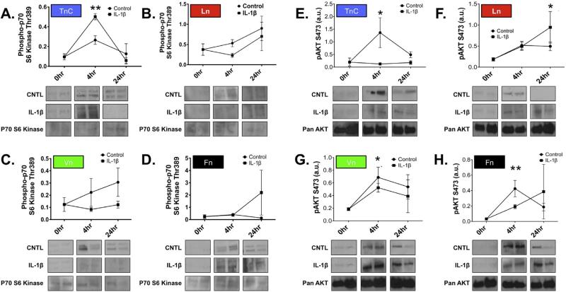
Fig. 3. The influence of ECM substrate on mTORC activation after scratch.
Time course analysis of S6 Kinase activation (pThr389; A–D) and Akt (pS473; E–H) following scratch wound in astrocytes grown on TnC (A and E), Ln (B and F), Vn (C and G), or Fn (D and H) with or without treatment with IL-1β. Representative immunoblots for activated S6 Kinase (pThr389) from each time point (as listed on the abscissa above each column) for Control (upper row) and IL-1β (lower row) for each ECM substrate, as indicated. Representative western blots for S6kinase from each time point show no significant change in overall levels of enzyme expression. (E–H) Representative western blots of time course analysis for pAkt activation (pS473) after scratch wound with or without treatment with IL-1β in primary astrocyte cultures. Immunoblotting for pan-Akt from each time point show no significant change in overall expression Akt expression over this time course. Quantification of western blots bands were corrected for sample loading relative to β-actin labeling of same blots (data presented as arbitrary units, a.u.) S6 Kinase data represent n = 8/ treatment/time point. (*P = 0.006) and pAkt data represent n = 8/ treatment/time point. (*P < 0.02, TnC, Ln; P < 0.04, Vn; **P < 0.005, Fn).

