. 2015 May 1;38(2):147–151. doi: 10.1590/S1415-4757382220140238

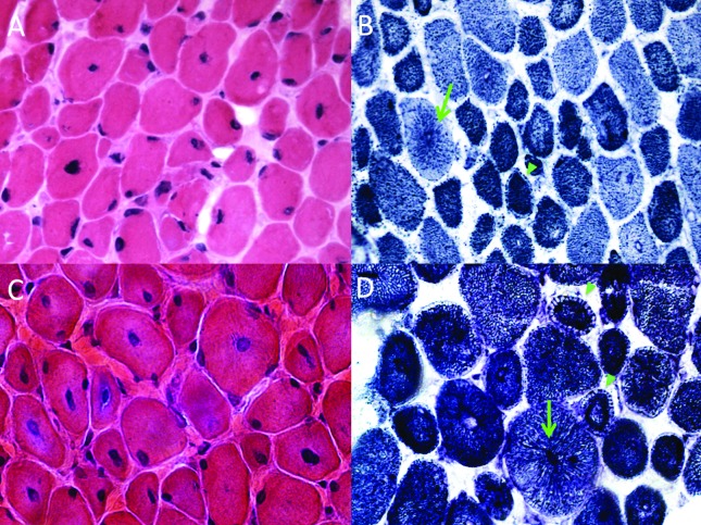
Figure 2. Muscle biopsy findings in the patients with DNM2 mutations. Patient 1: (A) histology showed moderate variability of muscle fiber size, fibers with prominence of centralized nuclei and mild increase of connective tissue (H&E stain, 40X); (B) fiber type architecture was abnormal, characterized, after oxidative stains, by fibers with radiating sarcoplasmic strands (aspect of “spoke of wheels”, green arrow), and ring-like fibers with a peripheral clear area (arrowheads) (NADH-TR, 40X). Patient 2: (C) as in Patient 1, the muscle biopsy showed a prominence of fibers with centralized nuclei and variable size, in addition to increased connective tissue (H&E stain, 40X); (D) oxidative stains also showed typical fibers with radiating sarcoplasmic strands (green arrow) and ring-like fibers (green arrowheads) (NADH-TR, 40X).

Figure 2