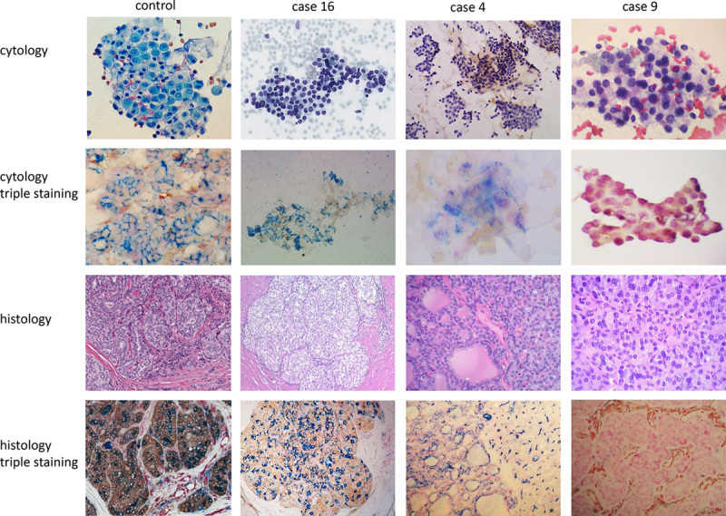
FIGURE 1.

Illustration of triple-stained cases. Case #16 showed very subtle nuclear clearing and slightly overcrowded nuclei on cytology (Diff Quik). This patient has been aspirated 3 times with 2 AUS and 1 FLUS cytology diagnoses. Triple staining on cytology revealed coexpression of Galectin-3 and HBME-1 in the same cell with no p27 labeling. The histology diagnosis is PTC. Case #4 showed mixed macrofollicles and microfollicles with very scant colloid on cytology (Diff Quik). The cytology diagnosis is FLUS. Triple staining on cytology demonstrated colabeling of p27 and HBME-1 in the same cell. Histology diagnosis is multiple nodular goiter. Control staining from liquid-based preparation and 1 PTC formalin-fixed paraffin-embedded section is shown in the first column. Case #9 showed microfollicles with very rare trabecular architecture on cytology (Pap stain). The cytology diagnosis is FLUS and histology diagnosis is follicular adenoma. Triple staining highlights the nuclei of benign follicular cells in both cytology and histology. The vascular stromal cells are positive for Galectin-3 on histology. AUS indicates atypia of undetermined significance; FLUS, follicular lesion of undetermined significance; PTC, papillary thyroid carcinoma.
