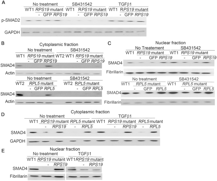
Fig 3. Change of SMAD4 after TGFβ inhibitor /activator treatment.
The iPSCs were cultured in iPSC medium, and treated with TGFβ inhibitor SB431542 (10μM) for 3 days or treated with TGFβ activator, TGFβ1 (20ng/ml) for 24h. DMSO treatment was used as the control. Protein levels were measured by Western blot analysis. A) Decrease of total p-SMAD2 after SB431542 treatment in all iPSCs, and increase of p-SMAD2 in both DBA iPSCs after the TGFβ1 treatment. B) Decrease of SMAD4 in the cytoplasmic fraction of all iPSCs after SB431542 treatment. C) Slight accumulation of SMAD4 in the nucleus after SB431542 treatment in DBA iPSCs with RPS19 or RPL5 mutations. D-E) Mild increase of cytoplasmic and nuclear SMAD4 in DBA iPSCs after the TGFβ1 treatment.

