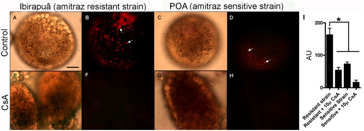
Fig 4. CsA-sensitive uptake of Sn-Protoporphyrin IX (Sn-Pp IX) is higher in amitraz-resistant ticks.

Digest cells from tick strains sensitive or resistant to amitraz were incubated in the presence of 100 μM Sn-Pp IX for 2 h. (A,B) Amitraz-resistant strain; (C,D) amitraz-sensitive strain; (E,F) amitraz-resistant strain preincubated with 10 μM CsA; (G,H) amitraz-sensitive strain preincubated with 10 μM CsA. A, C, E and G are DIC images. B, D, F and H are fluorescence images of the metalloporphyrin. Arrows indicate Sn-Pp IX fluorescence associated with hemosomes. The scale bar is 60 μm. (I) Quantitative analysis of Sn-Pp IX uptake measured as fluorescence intensity of digest cells from resistant and sensitive strains (expressed in arbitrary units; AU). Data shown are mean ± SEM from groups of 10 randomly chosen images obtained from three independent experiments; * means p < 0.001 (one-way ANOVA followed by Tukey’s test).
