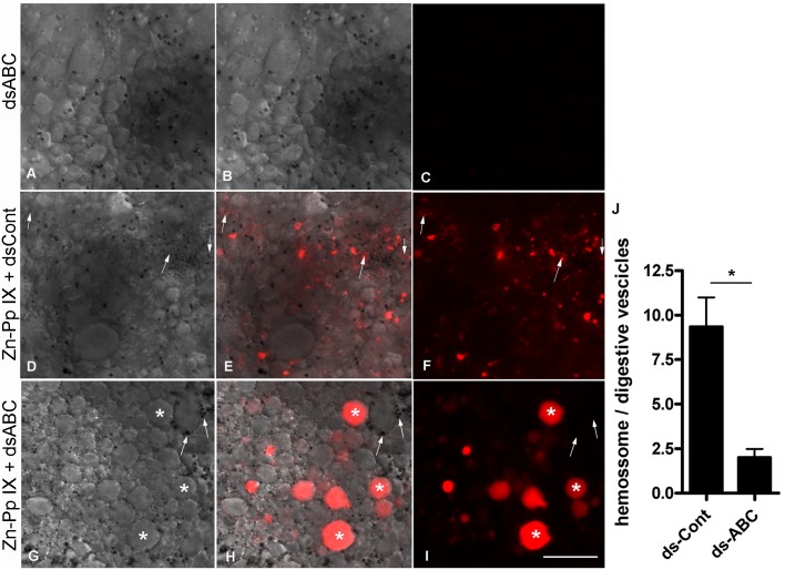
Fig 7. ABC transporter silencing impairs Zn-Pp IX traffic in digest cells.
Partially engorged females were collected from cattle and were artificially fed with blood supplemented with dsABC (A-C), with Zn-Pp IX plus dsCont (D-F) or with Zn-Pp IX plus dsABC (G-I). In all cases, the blood meal contained 0.5% DMSO (v/v). After 72 h ABM, digest cells were detached from the tissue, and differential interference contrast (DIC) (A, D and G) and Zn-Pp IX fluorescence images (C, F and I) were acquired. Merged images are shown in B, E and H. The white arrows indicate hemosomes (small vesicles) exhibiting a Zn-Pp IX signal in panels A_F; in panels G-I, some hemosomes are indicated, but with no fluorescence associated; white asterisks show digestive vesicles within the Zn-Pp IX signal. The scale bars are 20 μm in all images. The ratio of the number of Zn-PP positive hemosomes to digestive vesicles was measured in 15 randomly chosen images from each condition, obtained from three independent experiments (J). Data shown are mean ± SEM; * means p value < 0.002 (Student’s t test).

