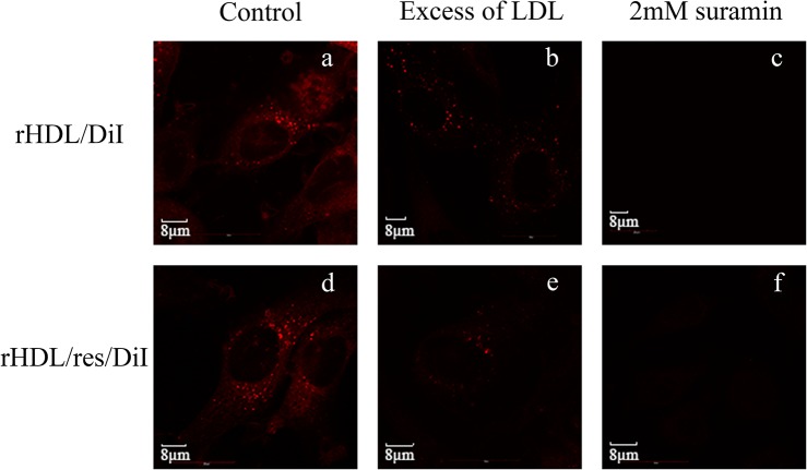
Fig 7. Confocal microscopy images of LDLr-mediated cellular uptake of rHDL/res.
Glioblastoma cells were treated with rHDL/DiI (Top) or rHDL/res/DiI (Bottom) in the absence (a and d) or the presence of 50-fold excess LDL (w/w) (b and e) or 2 mM suramin (c and f) at 37°C. for 3 h. The cells were visualized by the DiI fluorescence associated with lipids.

