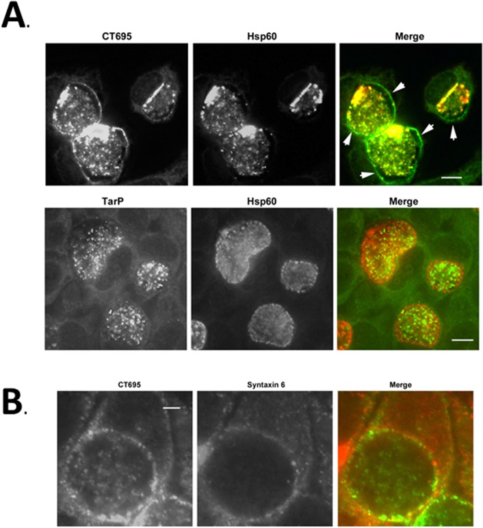
Fig 7. CT695 is secreted during late-cycle development and co-localizes with the chlamydial inclusion.
HeLa cells were infected with C. trachomatis L2 at an MOI of 1 and paraformaldehyde fixed at 24 hpi. (A). CT695 or TarP localization were assessed using α-CT695 or α-TarP, respectively. Chlamydiae were detected using α-Hsp60. Individual and merged channels of epifluorscence images are shown with specific detection of Chlamydia (red) and CT695 or TarP (green). Arrows indicate apparent inclusion membrane localization and scale bar = 10 μm. (B) HeLa cells were infected with C. trachomatis L2 at an MOI of 1 and paraformaldehyde fixed at 24 hpi. CT695 was detected with α-CT695 (green) wherease the position of the chlamydial inclusion membrane was visualized via staining with antibodies specific for Syntaxin-6 (red). Scale bar = 5 μm.

