Figure 3.

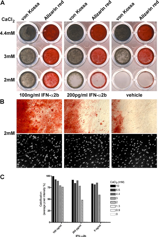
IFN-α enhances calcification in cultured hVSMCs. (A) hVSMCs were exposed to increasing concentrations of calcium (0, 0.9, 1.3, 2 [shown], 3 [shown], 4.4 [shown], 6.7, and 10 mmol/L CaCl2), with or without IFN-α2b (200 pg/mL or 100 ng/mL). Calcification was detected by von Kossa (gray-brown) or Alizarin red stain. IFN-α2b promoted hVSMC calcification particularly at 2 and 3 mmol/L calcium, with 100 ng/mL IFN-α2b having a stronger effect than 200 pg/mL IFN-α2b. Calcification was similar at calcium concentrations of 4.4 mmol/L or higher, with IFN-α2b having a small inducing effect. No calcification was detected at 1.3 mmol/L calcium concentration or lower, with or without IFN-α. Representative results are shown for three comparable experiments. (B) Hundred-times magnification pictures of the hVSMCs cultured with 2 mmol/L calcium, for the wells stained with Alizarin red (top) or for cell density with Hoechst (grayscale, bottom). (C) Calcification quantification of wells stained with von Kossa measured in average pixel intensity per well. An effect of IFN-α2b was particularly detected at 3 mmol/L CaCl2, with an intensity increase of 20% with 200 pg/mL or 100 ng/mL IFN-α2b compared to 0 ng/mL IFN-α2b, or at 2 mmol/L CaCl2, with an intensity increase of 48% with 200 pg/mL IFN-α2b or 76% with 100 ng/mL IFN-α2b, respectively. Representative results are shown for three comparable experiments. IFN, interferon; hVSMCs, human vascular smooth muscle cells.
