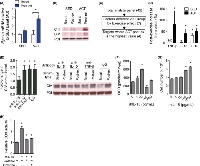
Fig 3.
IL-15 is responsible for exercise-stimulated mitochondrial biogenesis in skin in vitro. (A) Expression of Pgc-1α mRNA in buccal swabs prior to and immediately following a single session of aerobic exercise using Gapdh as a stable housekeeping gene. n = 8 per group. (B) Representative immunoblots of mitochondrial protein subunits of complex III (core 2, CIII) and complex IV (COX II, CIV) in primary dermal fibroblasts that were incubated for 48 h with media containing 10% human serum from the indicated blood sample. n = 4 replicates per condition. (C) Criteria used to analyze the results of a plasma cytokine panel for determination of factors induced by exercise that are likely to signal peripheral tissue mitochondrial biogenesis. Numbers in parentheses indicate the number of analytes that pertain to the successive criteria. (D) The percent change in plasma IL-15, IL-10, and TNF-β in SED and ACT groups in response to a single bout of exercise. n = 8 per group. (E) Immunoblots of mitochondrial complex IV protein in primary dermal fibroblasts incubated for 48 h in enriched media containing 10% basal or postexercise (Post-ex) serum from ACT subjects that was pretreated with control IgG, anti-TNF-β, anti-IL-15, or IL-10-neutralizing antibodies. n = 4 replicates per condition. (F) Oxygen consumption rate (OCR) and (G) cell counts of human primary dermal fibroblasts treated with the indicated concentrations of rhIL-15 for 48 h. n = 3. (H) COX activity in dermal fibroblasts that were untreated (CON) or incubated with 10 pg mL−1 rhIL-15 with and without 1 μm GW9662 or 5 μm pimozide for 48 h. Data are mean ± SE. Data in A and D were compared using a two-way repeated-measures anova. Data in E were compared using a paired t-test. Data in F–H were compared using a one-way anova. *P < 0.05 from basal or control condition. †P < 0.05 from all other conditions. #P < 0.05 from rhIL-15 only condition. ND, not detectable.

