Fig 4.

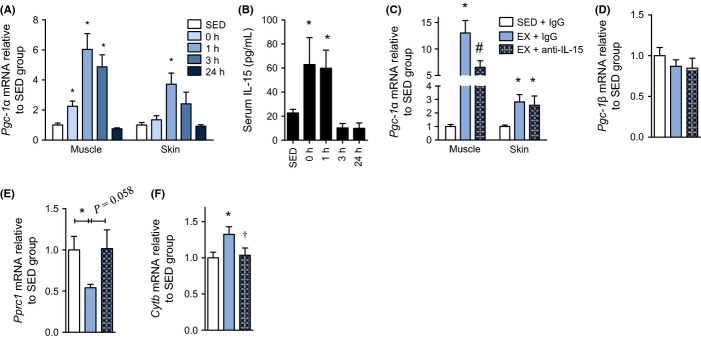
IL-15 partially regulates exercise-mediated mitochondrial signaling in skin and skeletal muscle. (A) qPCR of Pgc-1α mRNA in tissues from wild-type mice that were sacrificed at the indicated times after cessation of exercise or that did not exercise (SED). Muscle is quadriceps tissue, n = 9 per group. (B) Corresponding serum IL-15 concentration at the indicated times postexercise. n = 9 per group. (C) Pgc-1α mRNA expression in quadriceps muscle and skin from mice that remained sedentary or exercised in conjunction with injection of IgG control or anti-IL-15-neutralizing antibody. n = 6–10 per group. (D) Pgc-1β and (E) Pprc1 mRNA expression in skin tissue from mice that remained sedentary or exercised in conjunction with injection if IgG control or anti-IL-15-neutralizing antibody. n = 6–10 per group. (F) Cytb mRNA in skin acquired from wild-type mice that rested (SED) or were subjected to treadmill exercise in conjunction with tail vein injection of a total of 5 μg of IgG control or anti-IL-15 antibody. n = 6–10 per group. Mice in the neutralization experiments were injected with 2.5 μg antibody immediately before and after the exercise bout and sacrificed at 1 h following cessation of exercise. All acute mouse exercise experiments were performed at a 10-degree uphill grade at a speed of 16 m per minute for 1 h. mRNA expression analyses used β2-microglobulin as a stable housekeeping gene. Data are mean ± SE. Data were compared using a one-way anova. *P < 0.05 from SED group. †P < 0.05 from EX + IgG. #P < 0.05 from all other conditions.
