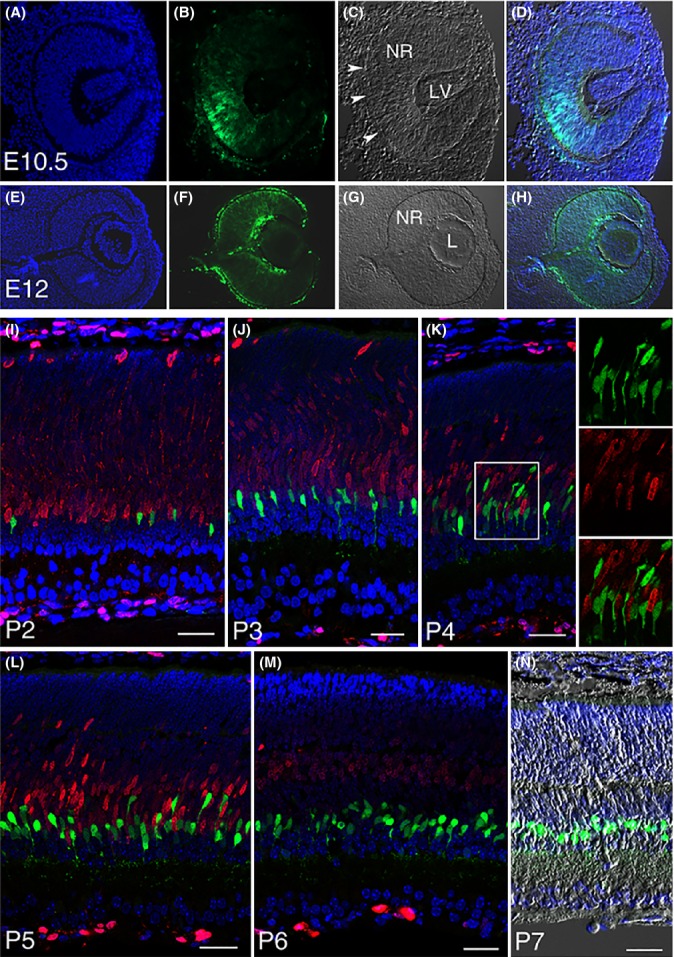
Fig 2.

Lgr5-EGFP expression at various stages of retinogenesis. (A–D) Lgr5-EGFP expression in the developing eye is first detected in the optic cup at E10.5, beginning in the middle of the optic cup and then expanding to its peripheral margin. Arrowheads in C highlight retinal pigmented epithelium. NR, neural retina; LV, lens vesicle. (E–H) Lgr5-EGFP expression in the neural retina (NR), lens (L), retinal pigmented epithelium, and surrounding mesenchymal cells at E12. (I–M) Confocal images of Lgr5EGFP-Ires-CreERT2 mouse retina co-stained with Ki67 (red) at postnatal day 2 through day 6. Lgr5-EGFP+ cells do not express Ki67 at any stage of late retinogenesis, indicating that they have exited the cell cycle. Boxed area in panel K is highlighted in the adjacent panels allowing for higher magnification views. (N) Merged image of DAPI staining, Lgr5-EGFP, and DIC of a retinal section at P7. In all relevant panels, Lgr5-EGFP is in green and DAPI staining is in blue. Scale bars = 30 μm.
