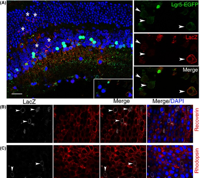
Fig 4.

Generation of new retinal cells in response to retinal injury. (A) Confocal images of a retinal cross section from Lgr5EGFP-Ires-CreERT2; Rosa26-LacZ mice analyzed with anti-β-galactosidase (LacZ) antibody (red) following retinal NMDA and growth factor injection. LacZ-positive cells are present in all three cell layers of the retina (indicated by stars in the inner nuclear layer and outer nuclear layer). The adjacent higher magnification views show co-localization of the Lgr5-EGFP signal with LacZ in cells of the loosely organized retinal ganglion layer (highlighted with arrowheads). (B) Co-staining of LacZ with recoverin (a marker for photoreceptors). Arrowhead marks LacZ and recoverin double-positive photoreceptor cells. (C) Co-staining of LacZ with rhodopsin (a marker for rod photoreceptors). Arrowheads mark LacZ and rhodopsin double-positive photoreceptor cells. Scale bars = 20 μm.
