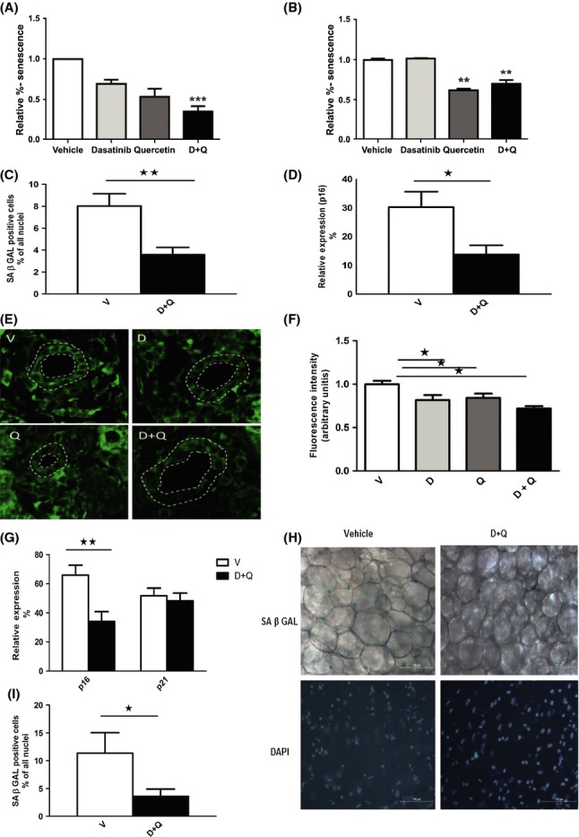
Fig 3.

Dasatinib and quercetin reduce senescent cell abundance in mice. (A) Effect of D (250 nm), Q (50 μm), or D+Q on levels of senescent Ercc1-deficient murine embryonic fibroblasts (MEFs). Cells were exposed to drugs for 48 h prior to analysis of SA-βGal+ cells using C12FDG. The data shown are means ± SEM of three replicates, ***P < 0.005; t-test. (B) Effect of D (500 nM), Q (100 μm), and D+Q on senescent bone marrow-derived mesenchymal stem cells (BM-MSCs) from progeroid Ercc1−/Δ mice. The senescent MSCs were exposed to the drugs for 48 h prior to analysis of SA-βGal activity. The data shown are means ± SEM of three replicates. **P < 0.001; anova. (C–D) The senescence markers, SA-βGal and p16, are reduced in inguinal fat of 24-month-old mice treated with a single dose of senolytics (D+Q) compared to vehicle only (V). Cellular SA-βGal activity assays and p16 expression by RT–PCR were carried out 5 days after treatment. N = 14; means ± SEM. **P < 0.002 for SA-βGal, *P < 0.01 for p16 (t-tests). (E–F) D+Q-treated mice have fewer liver p16+ cells than vehicle-treated mice. (E) Representative images of p16 mRNA FISH. Cholangiocytes are located between the white dotted lines that indicate the luminal and outer borders of bile canaliculi. (F) Semi-quantitative analysis of fluorescence intensity demonstrates decreased cholangiocyte p16 in drug-treated animals compared to vehicle. N = 8 animals per group. *P < 0.05; Mann–Whitney U-test. (G–I) Senolytic agents decrease p16 expression in quadricep muscles (G) and cellular SA-βGal in inguinal fat (H–I) of radiation-exposed mice. Mice with one leg exposed to 10 Gy radiation 3 months previously developed gray hair (Fig.5A) and senescent cell accumulation in the radiated leg. Mice were treated once with D+Q (solid bars) or vehicle (open bars). After 5 days, cellular SA-βGal activity and p16 mRNA were assayed in the radiated leg. N = 8; means ± SEM, p16: **P < 0.005; SA β-Gal: *P < 0.02; t-tests.
