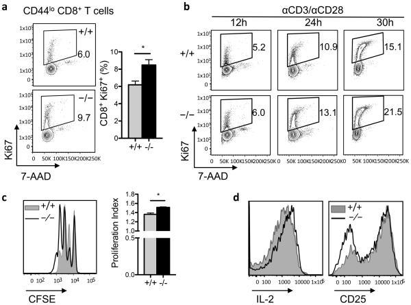
Figure 6. G0S2 restricts the proliferation of CD8+ T cells.
(a) Dual detection of the proliferation marker Ki67 and DNA content in naïve CD8+ T cells freshly isolated from the spleen of wild type and G0s2−/− mice. (b) Expression of the proliferation marker Ki67 and DNA content in CD8+ T cells cultured on plates coated with anti-CD3 antibody and anti-CD28 in the media. (c) Cell division of purified CD8+ T cells activated with anti-CD3/anti-CD28 was examined by dilution of the CFSE dye. Corresponding proliferation indexes are plotted as bar graphs (mean ± SD, n=3). (d) Cell surface expression of CD25 and intracellular IL-2 content in wild type and G0s2−/− CD8+ T cells activated in vitro.

