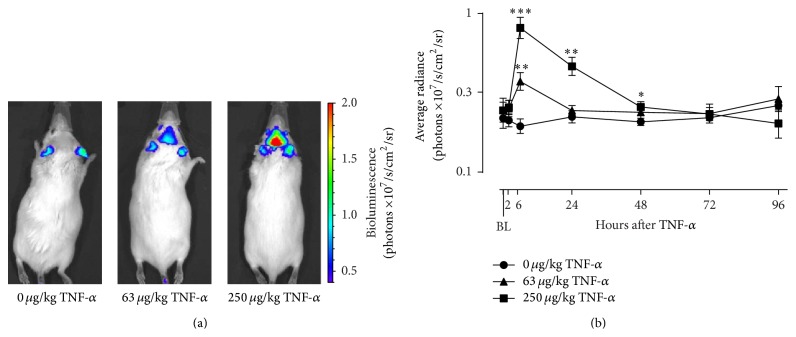
Figure 2.
TNF-α activates astrocytes in a dose- and time-dependent manner. Intraperitoneal injection of TNF-α caused a clear bioluminescent signal in the brain of Gfap-luc mice, as shown in representative images taken at 6 h after injection (a). This signal peaked at 6 h and then gradually waned over time (b). The color scale indicates the number of photons emitted from the animal per second. The graph is plotted as mean ± SEM (n = 7 per group). Data were analyzed by rmANOVA followed by independent samples t-test. BL: baseline. ∗ P < 0.05; ∗∗ P < 0.01; ∗∗∗ P < 0.001 compared to 0 μg/kg TNF-α.

