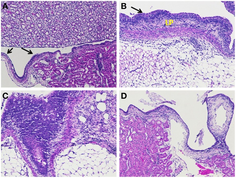
Figure 11.
Histological analyses of kidney tissue sections. (A) Kidney sections of the control group of rat showing normal transitional epithelium–urothelium (arrow) with no significant pathological changes. (B) Kidney section of diseased rat inoculated with S. aureus revealing moderate hyperplasia of the urothelium (arrow) and abundant lympho-plasmacytic infiltration (LP). (C) Kidney section of rat inoculated with S. aureus and treated with Gentamicin (LD) having moderate hyperplasia and abundant polymorphonuclear, few lympho-plasmacytic infiltrates beneath the urothelium. (D) Kidney section of rat inoculated with S. aureus and treated with Gentamicin (HD) having minimal infiltrates, predominantly lymphocytes (Gentamicin, LD, 8 mg/kg and HD, 50 mg/kg).

