
Figure 2.
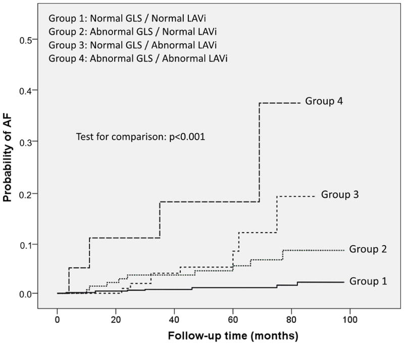
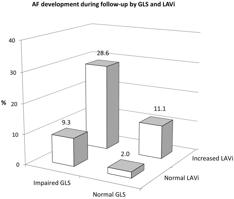
AF development during follow-up according to the combination of GLS and LAVi categories. A: Cumulative AF incidence in the four groups (p<0.001). B: Subjects with abnormal GLS and abnormal LAVi had an incidence of new-onset AF over 10-fold greater than the group with both normal parameters. Subjects with only one abnormality had an intermediate incidence of AF.