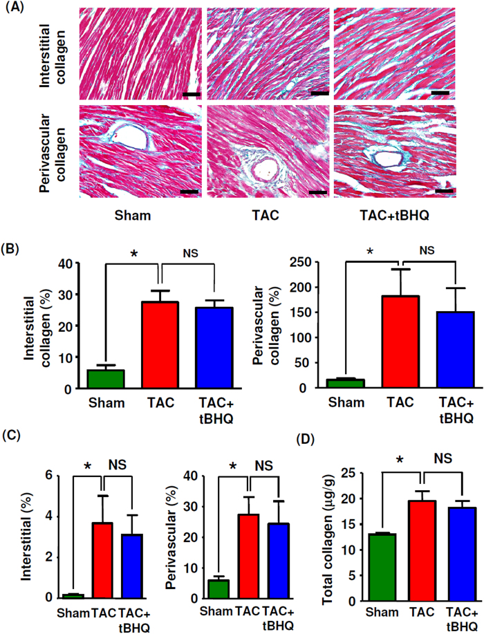
Figure 4. Effects of TBHQ on TAC-induced cardiac fibrosis.
(A) Masson’s trichrome staining showing interstitial and perivascular deposition of collagen fibers (blue color) (400 × images, bar = 50 μm). (B) Quantitative data of Masson’s trichrome staining for collagen deposition in LV (expressed as % area of the whole section for interstitial or as % area of the vessel lumen for perivascular) (n = 5–8 animals). (C) Quantitative data of picrosirius red staining for collagen deposition in LV (n = 5–8 animals). (D) LV collagen contents measured by hydroxyproline assay (n = 6 animals). *P < 0.05, one-way ANOVA. NS, no significance.

