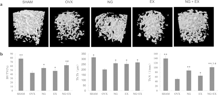
Figure 2.
(a) Representative sample from each group: 3D architecture of trabecular bone within the distal femoral metaphyseal region. (b) Effects of naringin or exercise on the trabecular bone volume, number of trabeculae, and thickness of the trabeculae of the distal femoral metaphysic in OVX rats by microtomography analysis. Values are means ± Standard deviation, n = 5. *p < 0.05, **p < 0.01 vs. OVX group; #p < 0.01 vs EX group; $p < 0.05 vs. NG group as evaluated by ANOVA.

