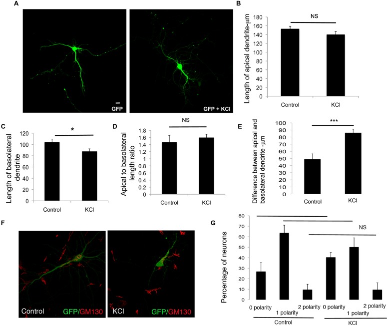
FIGURE 5.
Enhancing global neuronal activity suppresses net extension of basolateral dendrite extension in developing neurons. (A) Representative confocal images of neurons transfected with GFP and treated with or without KCl. (B) Length of apical dendrite. (C) Length of basolateral dendrite. (D) Ratio of lengths of apical to basolateral dendrites, and (E) Total dendritic length in neurons expressing GFP chronically treated without or with KCl at DIV 7. (Error bars represent SEM, Student’s t-test, P < 0.05, <0.005 -**, P < 0.005 -***, scale bar – 20 mm). (F) Confocal images of neurons expressing GFP treated without or with KCl and immunostained with anti-GM130. (G) Percentage of neurons with Golgi polarized toward 0, 1, or 2 dendrites in control or KCl treated neurons expressing GFP. (Error bars represent SEM, One way ANOVA, P < 0.05 -*, P < 0.0005 -***, scale bar – 20 microns).

