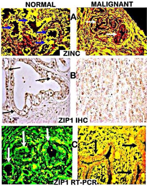
Figure 3.
Human normal prostate peripheral zone and prostate cancer tissue sections. A. Shows in situ zinc staining (yellow) in normal epithelium and loss of zinc (red) in early stage malignant acini cells. Arrows point to the acini glandular epithelium B. ZIP1 IHC shows (arrows) plasma membrane localized transporter in normal epithelium and absence in malignancy. C. In situ RT-PCR shows high gene expression (green stain) of ZIP1 in the normal glandular epithelium (arrows) and ZIP1 downregulation in malignancy. (Modified from [11]).

