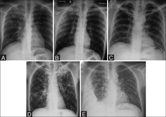
Figure 2 (A-E).

Chest radiographs in active TB. (A) CXR depicts RT upper zone consolidation with prominent RT hilum. (B) CXR in a different patient shows multiple coalescent air-space nodules in RT upper zone. (C) CXR in a different patient shows multiple ill-defined reticulo-nodular lesions in both lungs with basal predominance, suggestive of miliary TB. (D) CXR in a different patient shows active post-primary TB. Cavity with surrounding consolidation is seen in LT upper zone. Scattered air-space nodules are seen in both lungs with left hilar adenopathy. (E) There is RT-sided loculated pleural effusion with multiple air-space nodules scattered in both lungs
