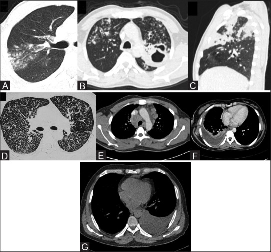
Figure 3 (A-G).

Chest CT in active TB. (A) Lung window (window center -600 HU, width 1200 HU) of chest CT depicts centrilobular and clustered nodules in posterior segment of RT upper lobe, suggesting active endobronchial infection. (B) Lung window of chest CT in a different patient shows cavity with surrounding consolidation in apicoposterior segment of LT upper lobe. Multiple centrilobular nodules with tree-in-bud branching pattern are also seen. (C) Sagittal multiplanar CT reformat lung window shows segmental consolidations in RT upper lobe. (D) Axial CT section lung window (in the same patient as in Figure 1c) in miliary TB shows multiple tiny discrete nodules randomly distributed in both lungs. (E) Axial CECT mediastinal window (window center 40 HU, width 400 HU) shows conglomerate rim-enhancing lymphadenopathy in paratracheal locations and multiple enlarged lymph nodes in prevascular location as well showing central necrosis. (F) Axial CECT mediastinal window shows RT-sided effusion with enhancing layers of pleura (split pleura sign) and rib crowding suggestive of empyema. (G) Axial non-contrast CT mediastinal window shows LT-sided free effusion
