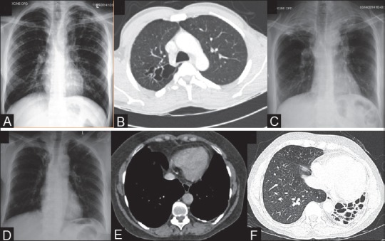
Figure 4(A-F).

Imaging features of healed TB. (A) CXR shows thin-walled cavity in left upper zone. Areas of fibro-bronchiectasis and fibrocalcific lesions are seen in left upper zone, RT upper and mid zones. (B) Axial CT lung window (window center -600 HU, width 1200 HU) shows clustered thin-walled cavities in superior segment RT lower lobe. (C) CXR shows volume loss in both upper zones with apical pleural thickening, pulled hila, fibro-bronchiectasis, and calcific foci. (D) CXR shows fibro-bronchiectasis both upper zones. (E) CECT mediastinal window (window center 40 HU, width 400 HU) shows left-sided pleural thickening and focal plaque-like calcifications. (F) CT lung window section in end-stage lung disease shows collapse and bronchiectasis involving the left lung with ipsilateral mediastinal shift and rib crowding
