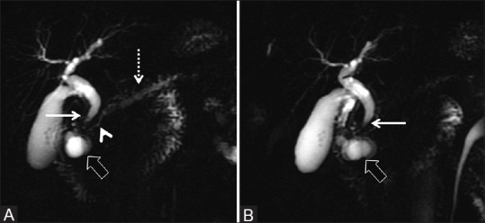
Figure 11(A and B).

(A and B) Thick slab 2D MRCP depicting characteristic features of paraduodenal pancreatitis as evidenced by paraduodenal cystic formations (thick arrow) with associated widening of the space between the descending duodenum, bile duct, and the pancreatic duct. There is associated smooth tapering of the distal bile duct (thin arrow). The pancreatic duct in the head region shows smooth decrease in diameter (arrowhead) with modest dilatation of the upstream duct (dotted arrow)
