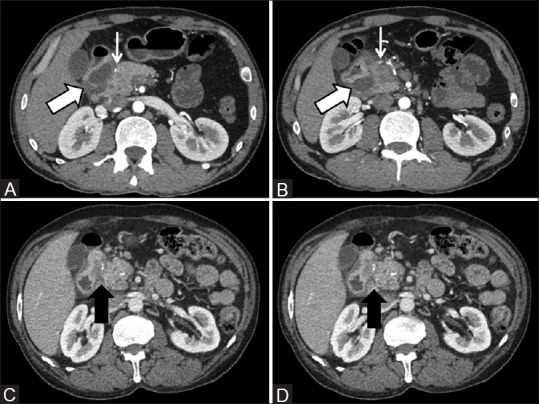
Figure 5(A-D).

(A and B) Cystic variant of paraduodenal pancreatitis displaying extensive cystic formations in the groove (thick arrow) causing medial displacement of the gastroduodenal artery which otherwise is normal in caliber (thin arrow) (C and D) Solid variant of paraduodenal pancreatitis wherein a hypoattenuating sheet-like fibrous tissue is seen within the pancreatico-duodenal groove (arrow)
