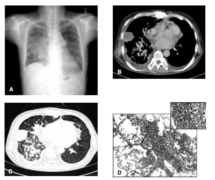
Figure 1.

A plain chest radiograph (A) and a computerized tomograph (B, C) of case 1, revealing multiple, variably sized nodular lesions in both upper lung fields. Multiple mediastinal lymphadenopathies are also shown in the right lobe paratracheal and subcarinal areas. Microscopic findings of a transthoracic lung biopsy show irregular thickening of the alveolar septa and perivascular connective tissue, due to the diffuse infiltration of atypical lymphoid cells, which were confirmed as being of B-cell lineage by immunohistochemical staining (D, × 100 & × 200, H-E).
