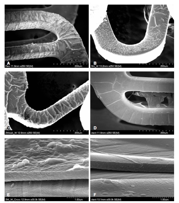
Figure 2.

Scanning electron microscopic findings of ReoPro® grafting after the washing test. The stent surface of the ReoPro® grafting; immediately (A), 5 hours (B), 2 weeks (C) and 4 weeks (D) after the washing test. E–F: Cross sections of the stent of the ReoPro® grafting, immediately (E) and 1 months (F) after the washing tests. Magnification. 250 in A–D and 30,000 in E–F.
