Figure 3.

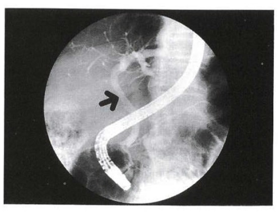
ERCP shows diffuse segmentally dilated Lt. IHD and slightly narrowed distal CHD (arrow). There is no filling defect in CBD and GB was invisible.

ERCP shows diffuse segmentally dilated Lt. IHD and slightly narrowed distal CHD (arrow). There is no filling defect in CBD and GB was invisible.