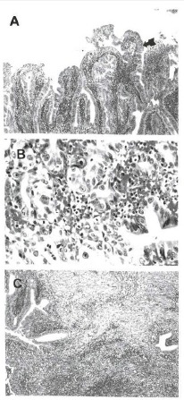
Figure 6.

GB biopsy findings. (A) Microscopy shows diffuse epithelial hyperplasia with partially high-grade dysplasia on GB neck (arrow) (H&E, ×40). (B) Microscopy shows focal areas of carcinomatous change on GB neck (H&E, ×100). (C) Microscopy shows acute nonspecific suppurative inflammation on GB fundus. (H&E, ×40).
