
Figure 1.
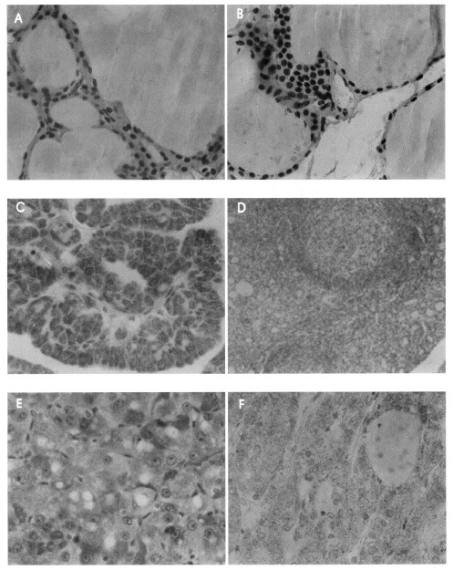
Immunohistochemical analysis of COX-2 in thyroid tissues. A: normal tissue (×400), B: adenomatous goiter (×400), C: papillary carcinoma (×400), D: Hashimoto’s thyroiditis (×100), E: follicular carcinoma (×400), F: follicular adenoma (×400).