Figure 13.

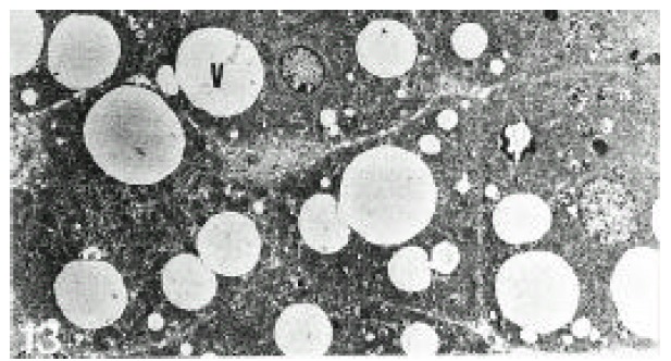
Electron micrograph of the liver in a patient with alcoholic fatty liver (case 4). Hepatocytes contained fat vacuoles of variable sizes. Fat vacuoles showed invagination into small ones (×3,332).

Electron micrograph of the liver in a patient with alcoholic fatty liver (case 4). Hepatocytes contained fat vacuoles of variable sizes. Fat vacuoles showed invagination into small ones (×3,332).