Figure 3.

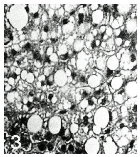
Higher magnification of the fatty liver from a patient with alcoholic fatty liver. Large fat vacuoles were distending hepatocytes and several fat vacuoles have coalesced (H & E, ×400).

Higher magnification of the fatty liver from a patient with alcoholic fatty liver. Large fat vacuoles were distending hepatocytes and several fat vacuoles have coalesced (H & E, ×400).