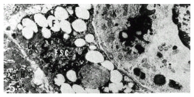
Figure 5.

Electron micrograph of a hepatic stellate cell (HSC) in alcoholic fatty liver (case 5). HSC contained fat droplets (F) of variable sizes. HSC was separated from hepatocyte (H), endothelial cells and Kupffer cell (K) by space of Disse (DS) containing collagen fibrils and numerous microvilli. Ce; centriole (×10,000).
