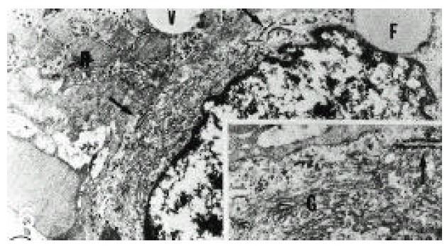
Figure 6.

Electron micrograph of hepatic stellate cell (HSC) from normal liver. HSC had well-developed Golgi apparatus and a few rough endoplasmic reticulum (arrows). H; hepatocyte, V; fat vacuole, F; fat droplet (×13,332).
Inset: higher magnification of a part of HSC showing well-developed Golgi apparatus (G), normally short, narrowed cisternae of endoplasmic reticulum (arrow) and glycogen granules in the cytoplasm. The Golgi apparatus consisted of several long and short, flattened cisternae and numerous vacuole (×26,664).
