Figure 1.

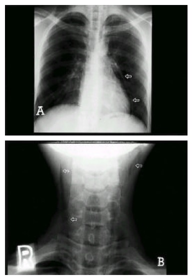
The radiograph of the chest (Panel A) showed pneumomediastinum and pneumopericardium (arrow) but it was clear on the lung fields. The radiograph of the neck (Panel B) showed subcutaneous emphysema (arrow).

The radiograph of the chest (Panel A) showed pneumomediastinum and pneumopericardium (arrow) but it was clear on the lung fields. The radiograph of the neck (Panel B) showed subcutaneous emphysema (arrow).