
Figure 1.
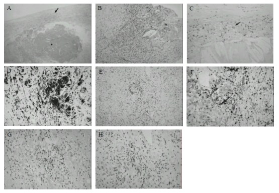
Histological analysis of human athereosclerotic plaques. (A) H&E staining of atherosclerotic plaque showing gross morphology. Lipid rich core (triangle) and fibrous cap with focal infiltration of immune cells (arrow) are indicated. (B) H&E staining of a region of heavy infiltration. (C) H&E staining showing a foam cell rich region (arrow). Immunohistochemical staining with anti-CD68 (D), anti-CD3 (E), anti-CD4 (F), anti-CD8 (G) and anti-TIA-1 (H) monoclonal antibodies. Magnification: (A), ×40; (B) and (E), ×100; (C), (D), (F), (G), and (H) ×200.