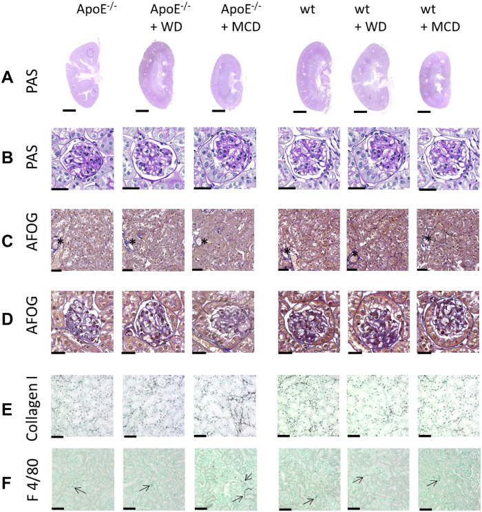
Figure 6. Kidney pathology in wt and ApoE−/− mice with or without WD or MCD diet.
Kidney morphology and detailed analyses of (A,B) Periodic Acid Schiff’s (PAS) and (C,D) Acidic Fuchsin Orange G (AFOG) stainings did not reveal any obvious pathological alterations in glomeruli (B,D), tubulointerstitium (C) or vessels (asterixes in C). No macroscopic differences were observed between the groups except of smaller kidney size in mice fed with MCD diet. (E) Immunohistochemistry for collagen type I, as a fibrosis marker and (F) F4/80, as a marker for renal monocyte/macrophage/dendritic cells showed normal findings comparable to wildtype mice fed with normal chow in all groups, apart from increased inflammation and fibrosis in ApoE−/− mice fed with MCD diet. Scale bars are 2.5 mm for A, 25 μm for B,D and 50 μm for C,E,F.

