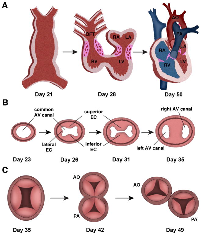
Fig. 1.
Cardiac valve development. A, At day 21 of gestation, the linear heart tube is formed and the primitive heart completes rightward looping by day 28 of human gestation. Significant remodeling of the inner curvature and growth of the ventricular chambers then occurs to result in the maturely developed heart with partitioned systemic (red) and pulmonary circulations (blue) by day 50. Regions of atrioventricular and outflow tract (OFT) cushion formation and development are shown in pink. LA left atrium, LV left ventricle, RA right atrium, RV right ventricle. B, Growth of the superior, inferior, and lateral endocardial cushions in the common atrioventricular canal is shown. Days of human gestation are noted. By day 35, the superior and inferior cushions have fused to divide to result in 2 atrioventricular openings. AV atrioventricular, EC endocardial cushions. C, Transverse section through the common outflow tract (truncus arteriosus) with 4 truncal swellings at day 35 of gestation. At day 42 of gestation, the common outflow tract has started to divide into the aorta (AO) and pulmonary artery (PA) and leaflet formation has begun. By day 49, the aorta and pulmonary artery are septated and each has 3 leaflets. Adapted from: Garg V. “Growth of the Normal Human Heart” In: Preedy VR, editor. Handbook of Growth and Growth Monitoring in Health and Disease. New York: Springer; 2011; and Garg V. “Insights into the Genetic Basis of Congenital Heart Disease”. Cell Mol Life Sci. 2006;63:1141–8 [4, 6]

